Traumatic Brain Injury
TRAUMATIC BRAIN INJURY
Road traffic accidents (RTA) are a leading cause of head /Brain injury and brain injury is the leading cause of death in RTA. Other causes of head injury include falls from height, falling heavy objects on the head, and assault. Early treatment is crucial to prevent secondary brain damage. The first hour is the Golden Hour where aggressive treatment can save the brain from lasting damage, which means the treatment should be started from the time the patient is transported from the accident site.
The Main Resuscitation Steps Include:
-
A
Clear & Protect Airway: Keep the airway clean from saliva, food particles, and vomitus to prevent hypoxia and aspiration.
-
B
Hemorrhage Control: Aggressively control active external bleeding and scalp hemorrhages to prevent hypovolemic collapse.
-
C
Maintain Hemodynamics: Maintain blood pressure and cerebral perfusion circulation to safeguard vulnerable brain tissue.
Severe head injuries with failing consciousness are managed in ICU preferably on ventilator for a couple of days.
CT Scan of the brain demonstrates the presence of haematoma, extent of brain contusion, displaced skull fracture and urgency for performing surgery.
THE GOLDEN HOUR IN NEUROTRAUMA INTERVENTION
Aggressive on-scene resuscitation, rapid diagnostic imaging, and prompt surgical decompression prevent irreversible secondary brain ischemic damage.
Causes
Falls are a common cause of TBIs, especially in older adults and young children, alongside high-velocity vehicular crashes and direct cranial trauma.
Symptoms
Headache, Confusion or disorientation, Temporary loss of consciousness, Nausea or vomiting, Fatigue & Difficulty sleeping.
Treatment
Stabilizing the patient, ensuring proper oxygen and blood flow, and addressing any life-threatening injuries through urgent surgical or medical protocols.
Rehabilitation
Physical therapy, occupational therapy, and speech therapy to address functional deficits and improve long-term quality of life.
Conditions Requiring Immediate Surgery
Prompt identification and rapid operative intervention are critical to preserving life and neurological function:
Extensive Scalp Laceration
This can lead to significant blood loss and hypotension. Immediate surgical exploration, wound debridement, and meticulous hemostatic closure are mandatory to prevent hemorrhagic shock.
Penetrating Injury & Depressed Fracture of the Skull
Such injuries carry a high risk of infection in the brain and hence need early attention. Surgery involves elevation of depressed bone fragments, dural repair, and removal of indriven debris to prevent meningoencephalitis and persistent seizure foci.
Extradural Haematoma
This is usually caused by a skull fracture injuring an underlying artery of the dura mater. This leads to rapid accumulation of blood clots, leading to acute compression of the brain, alteration of consciousness, and possible paralysis of limbs and coma. This life threatening condition needs evacuation of blood clots by ultra-urgent surgery. The results are very gratifying if performed in time. The patient who otherwise would have lost his/her life gains consciousness within hours.
Brain Contusion & Acute Subdural Haematoma
Severe injuries to the brain lead to contusion, laceration and accumulation of blood clots on the surface of the brain under the dura mater called subdural haematoma. This results in increase in intracranial pressure and rapid deterioration in consciousness or coma. The treatment is started to reduce intracranial pressure often by ventilatory support while some requiring immediate surgical intervention.
In this surgery, a large portion of the skull is lifted off and the dura mater is opened widely to allow the swollen brain to bulge outwards thus relieving the compressive effect from the brain stem. Results of this surgery are also gratifying provided the procedure is completed in time before any irreversible damage to the brain stem could take place.
Breakthrough Treatment Shows Promise in Traumatic Brain Injury
Multidisciplinary neuro-critical pathways designed to maximize cognitive and physical recovery:
Sports Injuries
Contact sports or activities with a risk of head impact can result in TBIs. Tailored concussion protocols ensure safe return-to-play while preventing second-impact syndrome.
Physical Examination
Assessing neurological function, pupillary reactivity, Glasgow Coma Scale (GCS) scoring, and subtle physical signs of cranial trauma.
Car Accidents
Motor vehicle accidents, including car, motorcycle, or bicycle accidents, can lead to traumatic brain injuries requiring swift polytrauma coordination.
Supportive Care
Providing ongoing care and assistance for individuals with persistent deficits, integrating neurocognitive monitoring and family counseling.
Clinical Radiographs & Traumatic Brain Injury Cases
Authentic diagnostic CT scans from Dr. Dilip Kiyawat's neurotrauma practice:
 Zoom
Zoom
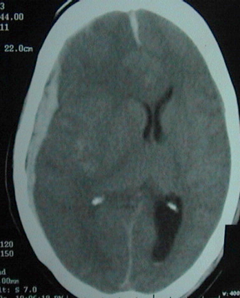
Acute Sub Dural Haematoma
Emergency CT brain demonstrating hyperdense crescentic extra-axial collection with severe subfalcine herniation requiring immediate decompressive craniectomy.
 Zoom
Zoom
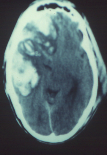
Cerebral Haemorrhagic Contusion
Axial non-contrast CT showing heterogeneous high-density contusions with extensive surrounding edema requiring intensive intracranial pressure management.
 Zoom
Zoom
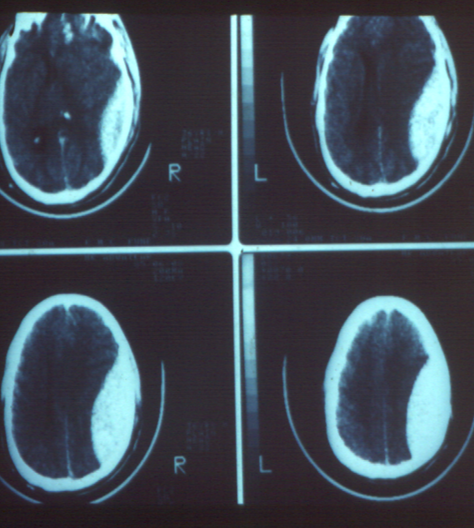
Extradural Haematoma
Classic biconvex, lentiform hyperdense extradural hematoma secondary to middle meningeal artery laceration, successfully treated with urgent craniotomy and evacuation.
For emergency neurotrauma consultation and appointment booking, you can reach Dr. Dilip Kiyawat at
-
Phone / 24/7 Emergency +91 98220 46043
-
Website drdilipkiyawatneurosurgeon.com